
Heel anatomy: what you need to know to understand your pain
The heel consists of the calcaneus, the Achilles tendon, the plantar fascia and a fat pad. Knowing its anatomy helps explain where heel pain comes from.
By Dr. Luigi ManziUpdated on
Does your heel hurt? Before starting any treatment, it's important to understand how it works. It may seem obvious, but I assure you that once you understand the anatomy of the heel, you can better understand why you're hurting and what you can do to feel better.
Over the years of clinical practice, I've noticed that patients who understand how their bodies work are the ones who heal faster. That's why today I want to explain everything you need to know about the structure of the heel.
What exactly is the heel?
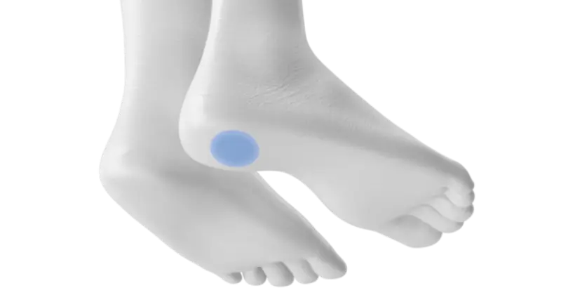
The heel isn't just the back of your foot. It's a complex structure. Incredibly complex, I'd say. Imagine it has to support your entire body weight every time you walk, run, or even just stand.
Its main function is to absorb shock when the foot hits the ground. But that's not all: the heel also helps maintain balance and provide the right push when we walk. That's why, when there's a problem in this area, all movement is affected.
The Heel Bones: The Calcaneus and Its Companions
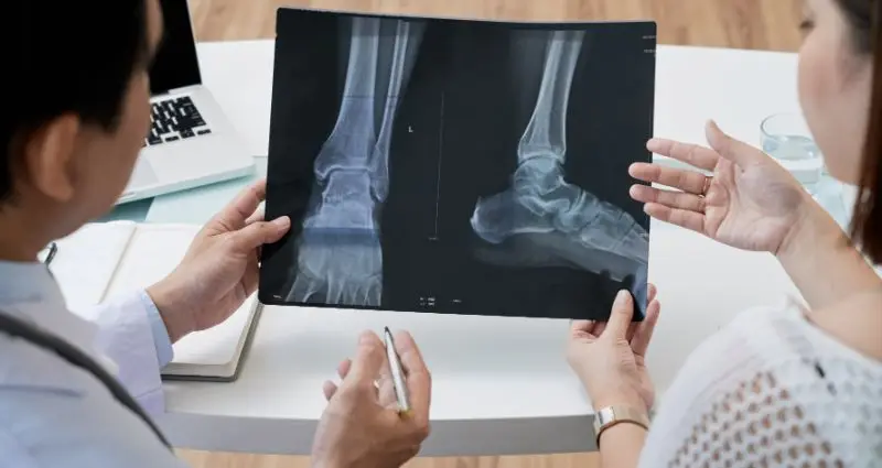
The calcaneus is the true protagonist. It is the largest bone in the foot and the one that primarily forms the heel. When we talk about calcaneus anatomy, we're referring to this parallelepiped-shaped structure located at the very back of the foot.
But it doesn't work alone. The anatomy of the calcaneus is closely linked to other heel bones, such as the cuboid, navicular, and cuneiforms. All these heel bones work together to create what we call the "arch of the foot."
The heel bone, also known as the calcaneus, has some interesting peculiarities. Its upper surface articulates with the talus (the bone above it), while the lower surface is where the muscles attach and where all of our body weight rests. This is why, when there is inflammation in this area, the pain can be truly intense.
The joints: where everything moves
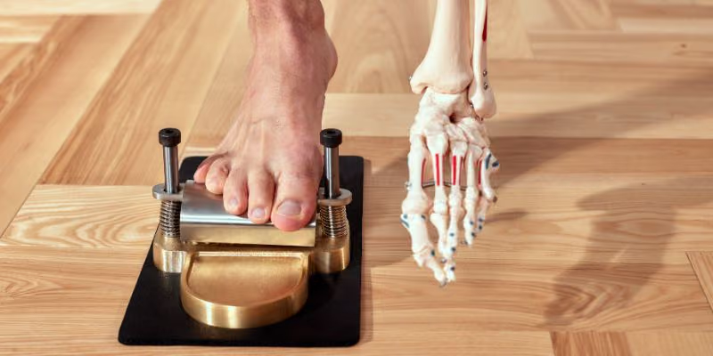
Now, this is an aspect we often overlook. The heel bone isn't a fixed block: it participates in various joints that allow the foot to move in all directions.
The talocalcaneal joint is the main one. It is here that the heel articulates with the talus, allowing for the inversion and eversion movements of the foot that we constantly use when walking on uneven terrain.
Then there's the calcaneocuboid joint, which connects the heel to the cuboid bone. And let's not forget the talonavicular and intertarsal joints: they all work synergistically to give the foot the mobility we often take for granted.
The muscles that move everything
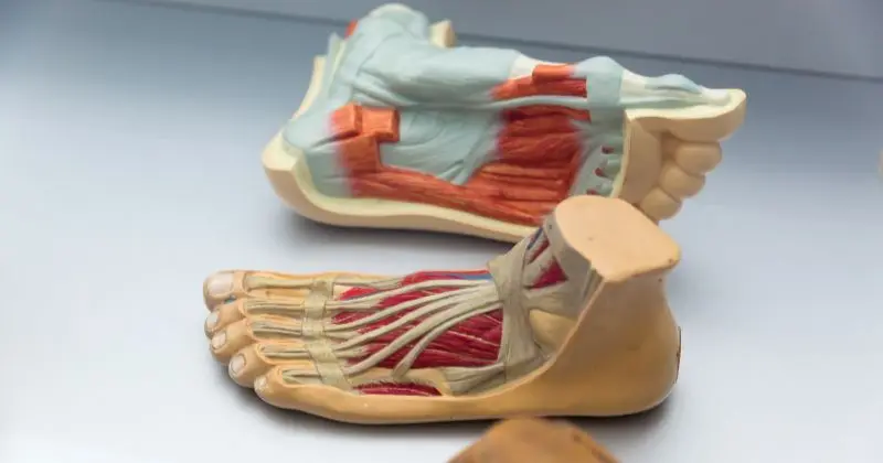
The triceps surae is the main muscle that acts on the heel. This muscle, composed of the gastrocnemius and soleus, inserts directly onto the heel via the Achilles tendon. Every time you walk, run, or even just stand on your toes, it's this muscle that does the work.
But there are also other important muscles: the tibialis posterior, the flexor digitorum longus, and the flexor hallucis longus. They all contribute to the movement and stability of the heel. When one of these muscles malfunctions, problems can begin.
Tendons: the “ropes” that transmit force
The Achilles tendon is undoubtedly the most famous. It's the largest and most powerful tendon in the body, connecting the calf muscles to the heel. When it becomes inflamed or ruptured, patients immediately recognize it: the pain is intense and function is compromised.
But there are also other tendons that pass around the heel: the plantaris tendon, the flexor digitorum longus, and the flexor hallucis longus. Each has its own specific role in the movement of the foot.
Nerves: the “powerhouse”
The heel is innervated primarily by the tibial nerve, which divides into the medial and lateral plantar nerves. The sural nerve also innervates the lateral aspect.
When these nerves become compressed or inflamed, the pain can be truly excruciating. I've seen patients describe the pain as "an electric shock" every time they put their foot down.
When things go wrong: the most common pathologies
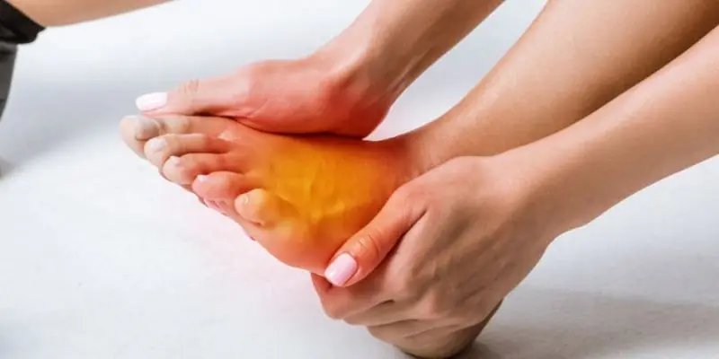
Plantar fasciitis is definitely the most common heel condition I see in my office. It's an inflammation of the plantar fascia that attaches to the heel. Pain typically occurs in the morning, when taking your first steps.
Heel spurs often accompany plantar fasciitis. They're a calcification that forms where the plantar fascia attaches to the bone. They don't always hurt, but when associated with inflammation, they can be very annoying.
Achilles tendinitis is another common condition. The tendon becomes inflamed, often due to overuse or poor posture. Heel bursitis, on the other hand, involves the bursae located between the tendon and the bone.
How do I diagnose?
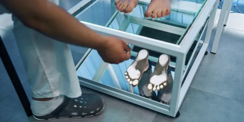
When a patient comes to me with heel pain, the first thing I do is a physical examination. I look at how they walk, check their foot mobility, and palpate the painful areas.
Then I ask about the symptoms: when does it hurt? In the morning? While walking? After sports? This information is crucial to guide the diagnosis.
If necessary, I order an X-ray to see the bones. In some cases, when I suspect soft tissue problems, an MRI may be helpful.
Treatments that really work
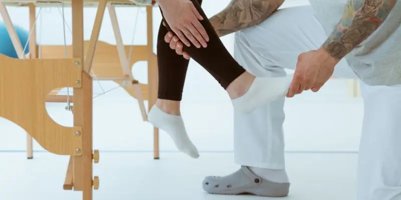
Conservative treatment is always the first choice. Relative rest, ice, and anti-inflammatories when necessary are recommended. Physical therapy is often very helpful, especially for stretching exercises.
In some cases, I use injections when the inflammation is particularly severe. Shockwave therapy can be effective for heel spurs and chronic plantar fasciitis.
I reserve surgical treatment for cases that don't respond to conservative therapy after at least six months of attempts. The procedure depends on the specific pathology, but we often achieve excellent results.
Prevention is better than cure
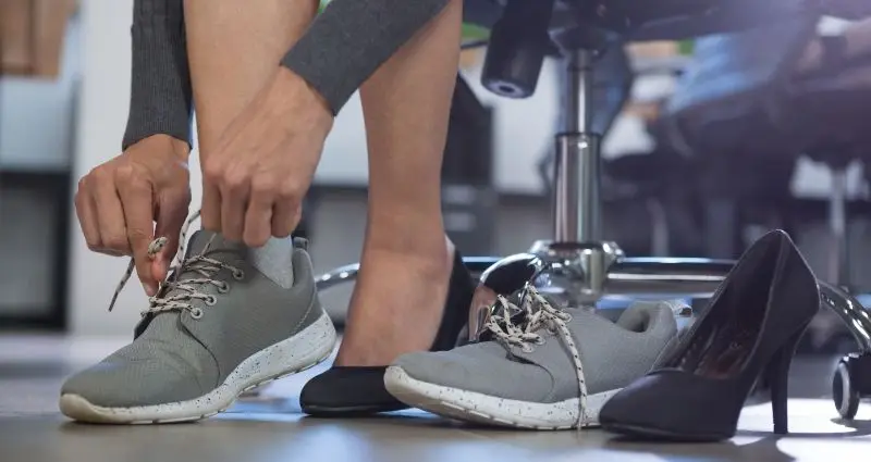
Wearing appropriate footwear is essential. Shoes with good arch support and a 2-3 cm heel are ideal for most people.
Daily stretching of the calf and plantar fascia helps tremendously. These simple exercises can make a difference. Maintaining a healthy body weight also reduces the strain on the heel.
SmartHallux: Where we can help you

Dr. Luigi Manzi, who has gained specific experience in this field, follows each patient with a personalized path using the most modern techniques.
The SmartHallux team treats conditions such as hallux valgus , Morton's neuroma , and other deformities that cause foot pain. Providing comprehensive support throughout the treatment process, SmartHallux can be a winning choice for effective treatment tailored to your needs. Book your specialist consultation with the SmartHallux team now (opens in a new tab).
FAQ
Why does my heel hurt more in the morning?
During the night, the plantar fascia shortens. When you put your foot down in the morning, it stretches abruptly, causing pain. After a few steps, the tissue "warms up" and the pain subsides.
Can I continue to play sports with heel pain?
It depends on the severity of the pain and the cause. Generally, it's best to reduce activity until we understand what's happening. Continuing to exercise can make the situation worse.
Are sneakers good for heel pain?
Not all. They must have good arch support and adequate cushioning. I often recommend using specific insoles.
How long does it take to heal?
It varies greatly depending on the condition. Plantar fasciitis can resolve in a few weeks with the right treatment, but in some cases it can take months. The key is to be consistent with treatment.